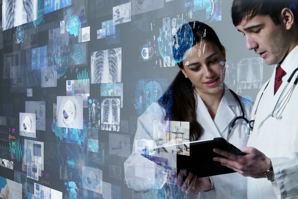
Integrating advanced technologies into the image interpretation pipeline can help the healthcare sector address challenges associated with large amounts of data. Custom software development of medical image analysis tools is a critical step in this process.
Over the past few decades, medical imaging has evolved from relatively primitive 2D scans into high-resolution sources of valuable information. Just imagine: as early as 2016, the volume of medical image data already accounted for a striking 90% of all healthcare big data. Given that interpreting such pictures is a time-consuming process, this has resulted in a dramatic shortage of radiologists.
Read also: Guide to Developing Healthcare Analytics Software
In this article, we’ll talk about the potential of image analysis tools for various healthcare departments and the industry as a whole. In addition, we’ll highlight the key points of medical image analysis software development.
How medical image analysis software can impact healthcare
As the name suggests, medical image analysis solutions can either interpret or help clinicians to interpret information obtained from radiology exams. These include, but aren’t limited to, computed tomography (CT), magnetic resonance imaging (MRI), thermography, or echocardiography scans. The increasing number of solutions for medical image analysis leverage machine learning (ML), deep learning, and big data analytics in order to:
-
Identify the exact treatment area and thus prevent (or minimize) possible damage to healthy tissues
-
Find pathologies even in images that don’t reveal any visible abnormalities
-
Sift through hundreds of scans, ‘flag’ images with abnormalities, and prioritize them based on complexity, allowing doctors to focus on severe cases
-
Validate diagnosis
-
Monitor disease progression by comparing images made at different points in time
-
Streamline communication and image data sharing between various decision-makers
Due to the above capabilities, radiology image analysis software can minimize clinical errors, avoid unnecessary procedures, reduce radiation exposure, facilitate early diagnostics and thus increase recovery/survival rates, accelerate the image examination workflow, and minimize hospital stays. As a result, healthcare providers can save their time and resources while improving the quality of care.
Medical image analysis software: top market trends
Despite the decline in the number of imaging sessions due to COVID-19, the growth rate of the medical image analysis market remains stable. According to a study released by Mordor Intelligence in 2021, this market is projected to grow at a CAGR of 8.1%, which is no different from estimates by Grand View Research based on data from 2019. But most importantly, it’s isn’t just growing. It’s changing, too. Let’s elaborate on this below.
AI adoption
Machine learning, deep learning, and computer vision are the key technologies in modern radiology. And this is more than just an assumption. Based on the data from 2020, the world market for AI medical imaging solutions is estimated to grow at a CAGR of a whopping 26%, as Signify Research suggests in their report. The study further explains that such tremendous growth owes to:
-
The record number of AI solutions approved by the FDA in 2020
-
New medical billing codes for AI-aided imaging
-
The first Class III NMPA approvals in China that permit the use of AI for diagnostics
And there’s nothing surprising in these positive developments. After all, AI has the potential to solve most of the pain points in the medical imaging niche. Capable of analyzing large amounts of imaging data and “seeing” details that remain invisible to the human eye, AI technologies can automate the image analysis workflow, decrease the number of wrong or missed diagnoses, reduce the need for invasive diagnostic tests, and beyond.
A shift to 3D
Two-dimensional imaging is still the backbone of the radiology industry. Due to the high resolution and low cost, it accounted for 71% of the market in 2020. Still, 2D has its drawbacks: by imposing a 3D structure on a 2D plate, you risk missing important details. As a result, hospitals and other healthcare organizations are increasingly turning to 3D to make up for these limitations.
For example, in breast imaging, digital tomosynthesis (3D mammography) has already become the “gold standard”. Since both cancer tissue and fibroglandular (the milk glands and ducts in breasts) appear white on a mammogram, they can overlap on a 2D image, which might prevent a healthcare provider from identifying the abnormality in time. On the contrary, tomosynthesis allows viewing an area of interest from different angles. Thus, 3D imaging increases the accuracy of anomaly detection, especially when combined with AI, which is able to process high volumes of data.
Since MRI, CT, and X-Ray scanners are evolving to produce three-dimensional scans of higher quality, the demand for analysis software with the ability to process 3D images will only increase.
Cloud computing
As radiology devices are becoming more sophisticated, the amount of data contained in medical images increases. This is also due to the fact that scanners typically produce tons of images per patient. All this information must be stored somewhere for further analysis.
To handle this issue, healthcare providers are increasingly switching to the cloud. A prime example of how you can benefit from cloud architecture as a healthcare facility or a medical image analysis software provider is Aidoc. Built on AWS, this AI solution from the healthtech provider has already analyzed over three million medical images at more than 300 healthcare facilities worldwide. Apart from enabling Aidoc’s clients to process enormous quantities of information in a safe environment, the cloud architecture allows the AI solution provider to release new algorithms every three months.
On top of that, cloud computing also facilitates medical image data sharing, which is particularly important in the COVID-19 era.
Integrated software
The increasing cloud adoption is also a result of the rising demand for integrated systems for medical image analysis: in fact, such solutions dominate the medical image analysis market with a revenue share of 55.5%. A single toolkit designed for a host of radiology operations (such as image acquisition, processing, analysis, and management) and a centralized healthcare data storage with live access to multiple users is a much more convenient and cost-effective option than a variety of standalone tools. That’s why if you are planning to develop medical image analysis software, be sure that it seamlessly integrates with your other radiology tools.
Medical image analysis: areas of application
Any healthcare department that uses medical imaging for diagnosis can benefit from medical image analysis software. Let’s take a look at a few examples.
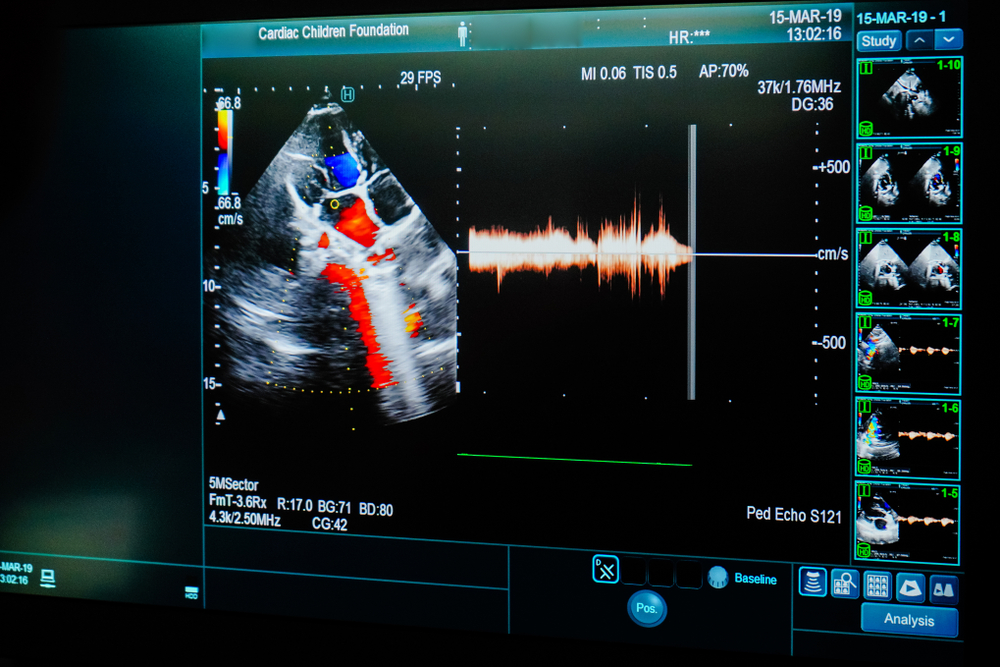
Cardiology
In 2019, cardiology accounted for 20% of the radiology image analysis market. And with cardiovascular diseases remaining the leading cause of death, this trend isn’t going anywhere. A closer look at this market will reveal that advanced algorithms are utilized in various stages of cardiovascular image analysis. For example, Arterys Cardio AI, a well-known MRI solution, automates routine tasks in a cardiac analysis workflow, including 2D visualization, 3D and 4D reconstruction, semi-quantitative perfusion, quantitative delayed enhancement, and tissue characterization. The solution providers promise that Arterys can save you up to 25 minutes per study.
Orthopedics
In orthopedics, ML algorithms take 3D reconstruction to the next level. For example, when a patient has a bone or limb fracture, an ML-driven 3D modeling solution can automatically align bone parts, thus avoiding minor segment misalignment typical of manual reconstruction. The resulting image can serve as a critical basis for further treatment, including surgery or implant design. Besides, due to their powerful image segmentation capabilities, medical image analysis solutions for orthopedics can be efficient for the detection of osteosarcoma, a type of bone tumor.
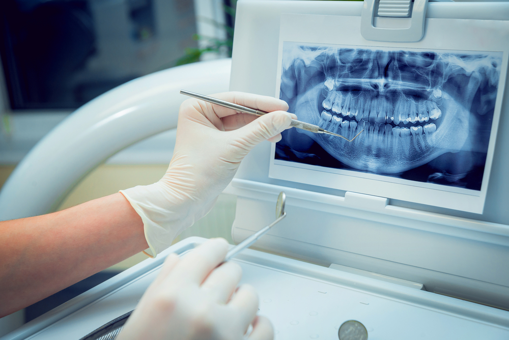
Dentistry
Dentistry is one of the most challenging areas when it comes to image-based decision-making: the oral cavity is densely cluttered with tiny structures that often overlap. Besides, teeth typically vary in size, shape, and position creating an additional obstacle for analysis. To ensure maximum diagnosis accuracy and workflow speed, dentists and radiologists use medical image analysis software. These tools are particularly useful for detecting caries, osteoporosis, and other pathologies.
Apart from abnormality detection, image analysis tools (or computer-aided design and computer-aided manufacturing systems, to be more precise) are also used for designing orthodontic appliances and dental prostheses.
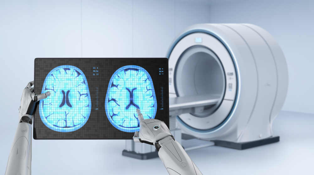
Neurology
In brain imaging, AI-driven analysis solutions assist clinicians in detecting pathologies that are otherwise invisible to the human eye. For example, a group of researchers from the University of California revealed that AI is capable of identifying the early signs of Alzheimer’s. They built an ML model able to identify the disease by “viewing” brain images taken six years before the patients were diagnosed with the condition.
Another example is a clinical study conducted by Canadian epilepsy experts. They managed to train an ML model to analyze epilepsy patients’ brain scans that don't reveal any visible signs of the condition. As a result, the solution helped them to locate and remove the source of seizures in a 27-year-old woman.
Breast oncology
With one in eight women having the potential to develop breast cancer at some point in her life, timely tumor detection remains one of the biggest concerns in the breast imaging domain. Fortunately, many advances have been made in this respect. For example, AI models have proven to be capable of identifying tumors that are otherwise impossible to detect. Meanwhile, according to NCBI, deep-learning models are capable of performing at the same level as radiologists in terms of diagnosis accuracy during breast cancer screening.
The main medical image analysis software features
If you take a closer look at today’s medical image software market, you’ll find a large array of solutions that vary in complexity, which makes identifying core functionalities an arduous task. Still, based on our experience, computer-aided medical image analysis isn’t efficient without the following features:
-
Image quality improvement
-
Image segmentation
-
Image registration
-
Quantification
-
3D reconstruction and 2D visualization
So, take these into consideration before developing medical image processing software. Now, let’s discuss each of these functionalities in more detail.
Image quality improvement
Since poor image quality can make important details hard to discern and thus put a patient’s well-being at risk, image enhancement is crucial for further analysis. It usually includes techniques like noise reduction, contrast enhancement, spatial aliasing compensation, artifacts removal, and so on.
Image segmentation
Segmentation is the procedure of dividing a medical picture into separate parts, such as organs, bones, tissues, or blood vessels. Besides, this process might also imply detection of pathologies in the region of interest, such as tumors, nodules, and other abnormalities.
Image registration
Image registration is the process of pulling data from multiple images into a single image. It falls into two subcategories:
-
In image fusion, data comes from different sources. For example, in order to learn how the patient’s anatomy correlates with their metabolism, clinicians can fuse CT scans with PET images.
-
4D fusion implies aligning images taken at different points in time. This technique is applied to monitor both long-term changes (such as a lesion’s growth over several years) or changes within the same imaging sessions (for example, to monitor a respiratory function).
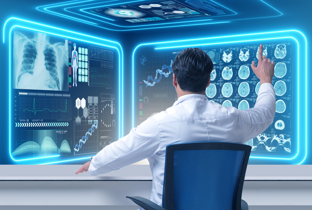
3D reconstruction and 2D visualization
3D reconstruction is a post-processing technique that implies combining multiple 2D images that depict the same area of interest from different angles into a single image. Able to view a single area of interest in 3D, clinicians can interpret abnormalities more efficiently. 3D reconstruction is opposed to 2D visualization, a process of dividing a 3D visualization into 2D components or segmenting a 2D image into smaller sections for greater detail.
Quantification
Quantification is the process of associating image segments with diagnostic information, such as their form, size, texture, morphology, and changes over time. It’s essential to consider this feature when it comes to building custom medical image analysis solutions.
The key challenges of custom medical image analysis software development

Today’s radiology scan analysis tools play a significant role in improving the entire medical imaging analysis workflow and increasing the number of positive patient outcomes.
However, neither you (if you are a healthcare provider) nor your clients (if you are a healthtech service provider) will be able to enjoy the full potential of modern technologies if you overlook challenges associated with the implementation of medical image analysis and processing software. Below are some of them:
-
Security and compliance. Medical imaging involves collecting patient identifiable information, which is protected by HIPAA. Since the HIPAA penalties can reach millions of dollars (to say nothing of ruined reputation), compliance is crucial. Make sure that your tech stack includes technologies that respect all necessary standards and that your data is anonymized once it’s outside your premises.
-
AI model training. The main limitation of algorithmic models is that they will do what you train them to do. That’s why it’s important to choose only applicable algorithms and train them on high-quality data sets. Be sure that you use correct, consistent, and complete data to feed your algorithmic models. Otherwise, they will give inaccurate results.
-
Training data. It’s impossible to build medical image analysis software that facilitates clinical decision-making without large volumes of data. Given that, be sure that before the development begins, you know exactly where and how you’re going to collect your training data, how they’ll be prepared, and if there’s secure storage with enough space.
-
Multimodality issues. Given the potential of image fusion, there’s an increasing demand for multimodality imaging platforms capable of processing and aligning images of several modalities, such as CT, MRI, X-ray, or ultrasound. This might be a daunting task for your development team.
The development of medical imaging analysis software requires relevant tech expertise, as well as niche experience. To make up for the lack of the required professionals in-house, companies increasingly enter into partnerships with other businesses. However, if you need to set the ball rolling and build a product as soon as possible, feel free to outsource development to a trusted vendor, like Demigos.
Conclusion
Since there’s still a lot of room for improvement in medical image analysis, the competition in this market is becoming fierce. That said, as a software provider, you need to stay attuned to the pulse of the ever-changing healthtech market trends and remain aware of possible difficulties that you might experience during the implementation stages.
At Demigos, we can help you with this. And in addition to explaining how to create custom medical image analysis solutions that bring value to their users, we can also build them for you—just reach out to us.